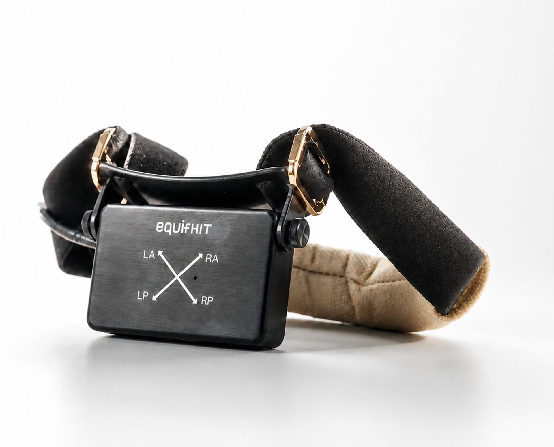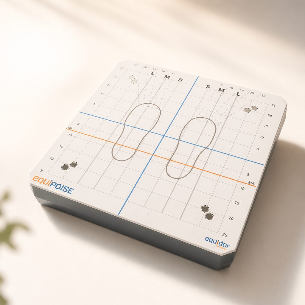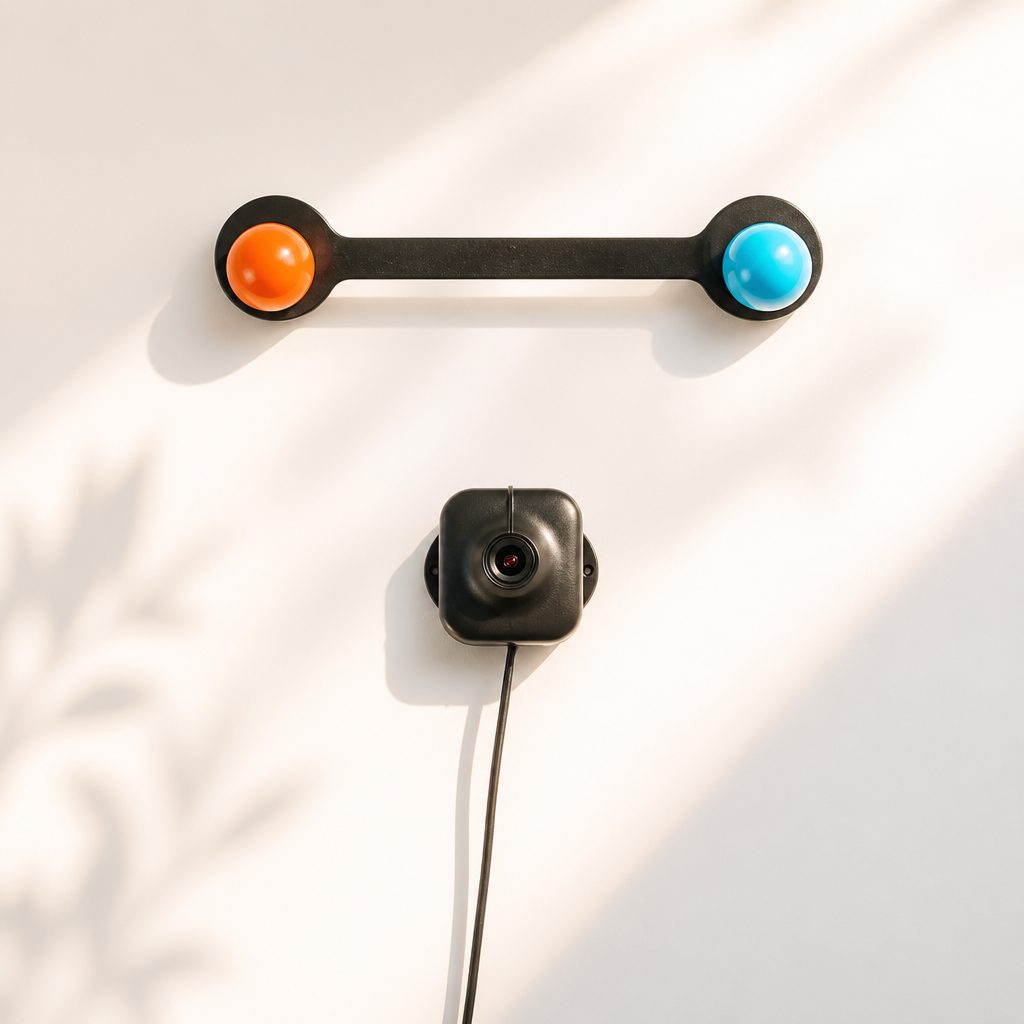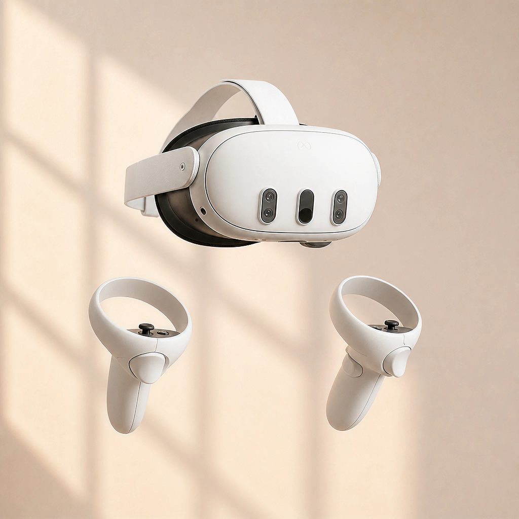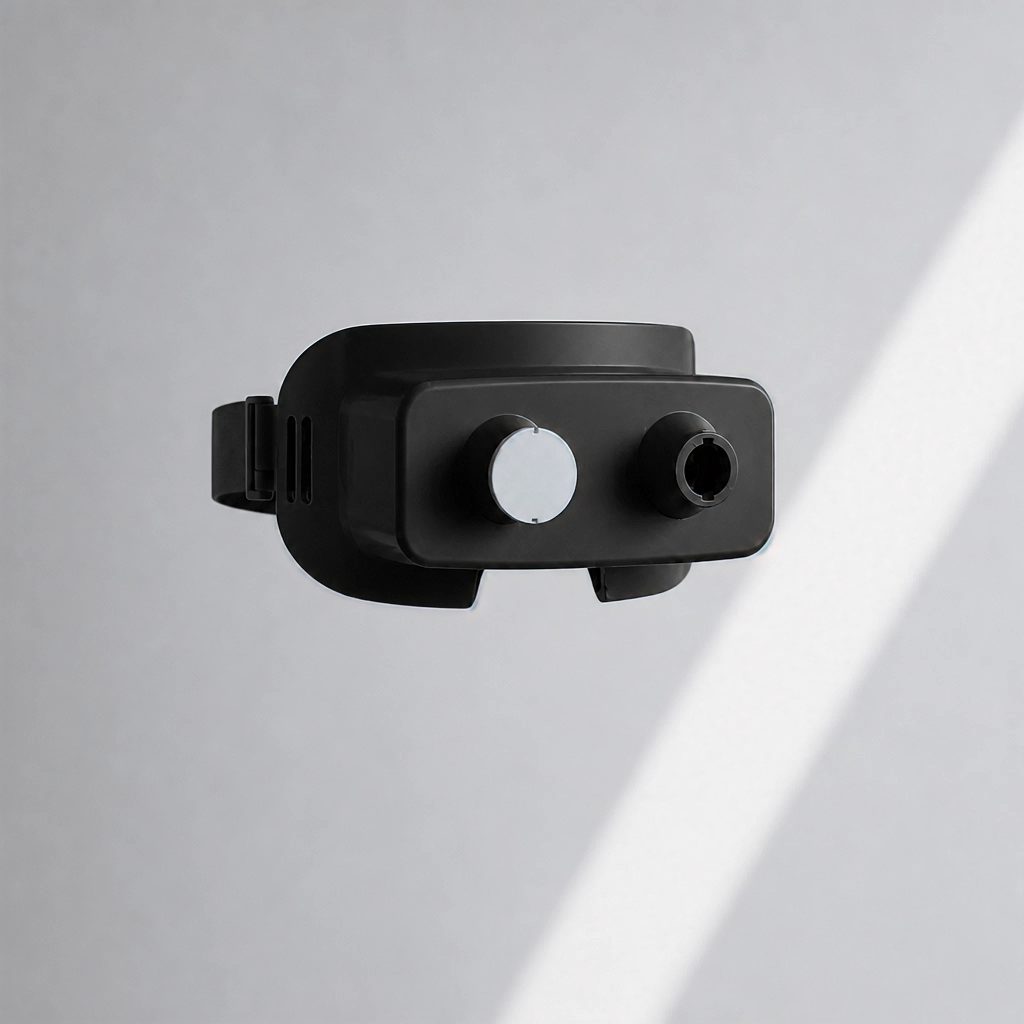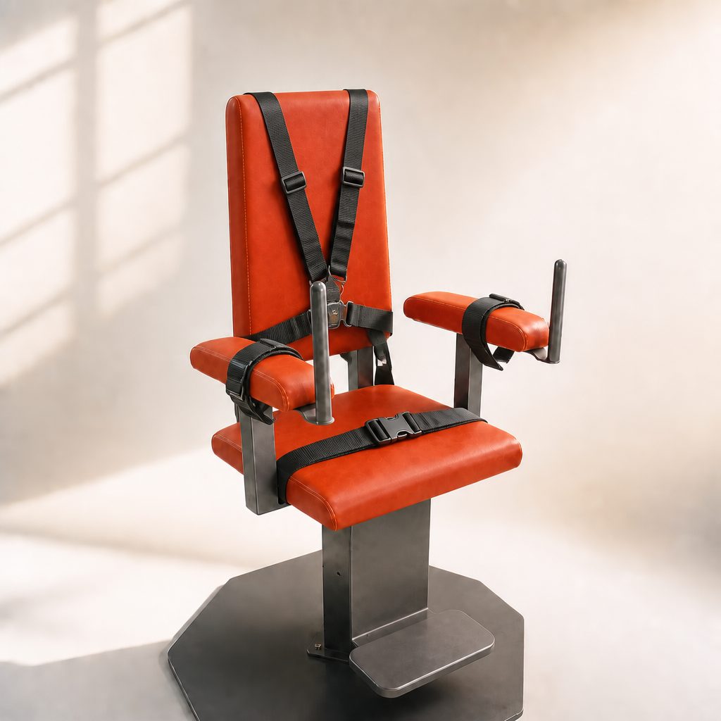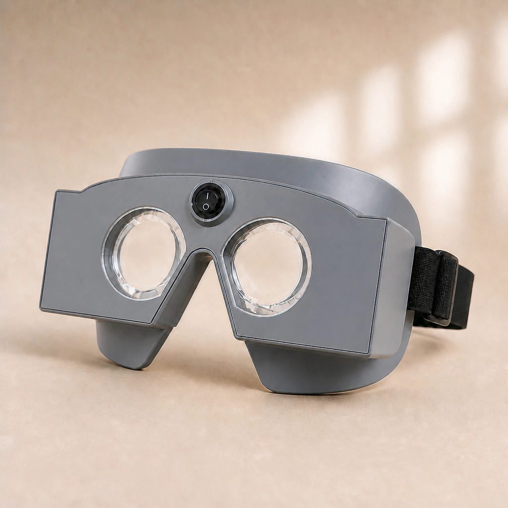

Head-mounted eye and head tracking system for comprehensive videonystagmography and oculomotor assessment.

The vestibulo-ocular reflex evolved to stabilise gaze against head movement, so interpreting nystagmus without simultaneous head data leaves the clinical picture incomplete. Even healthy individuals exhibit transient per-positioning nystagmus during position changes — easily misread as pathology when the eyes are studied in isolation. equiCOG, true to its name (cranio-oculography), captures eye behaviour in the context of head behaviour, giving every recording the reference frame it needs for accurate interpretation and confident decision-making.
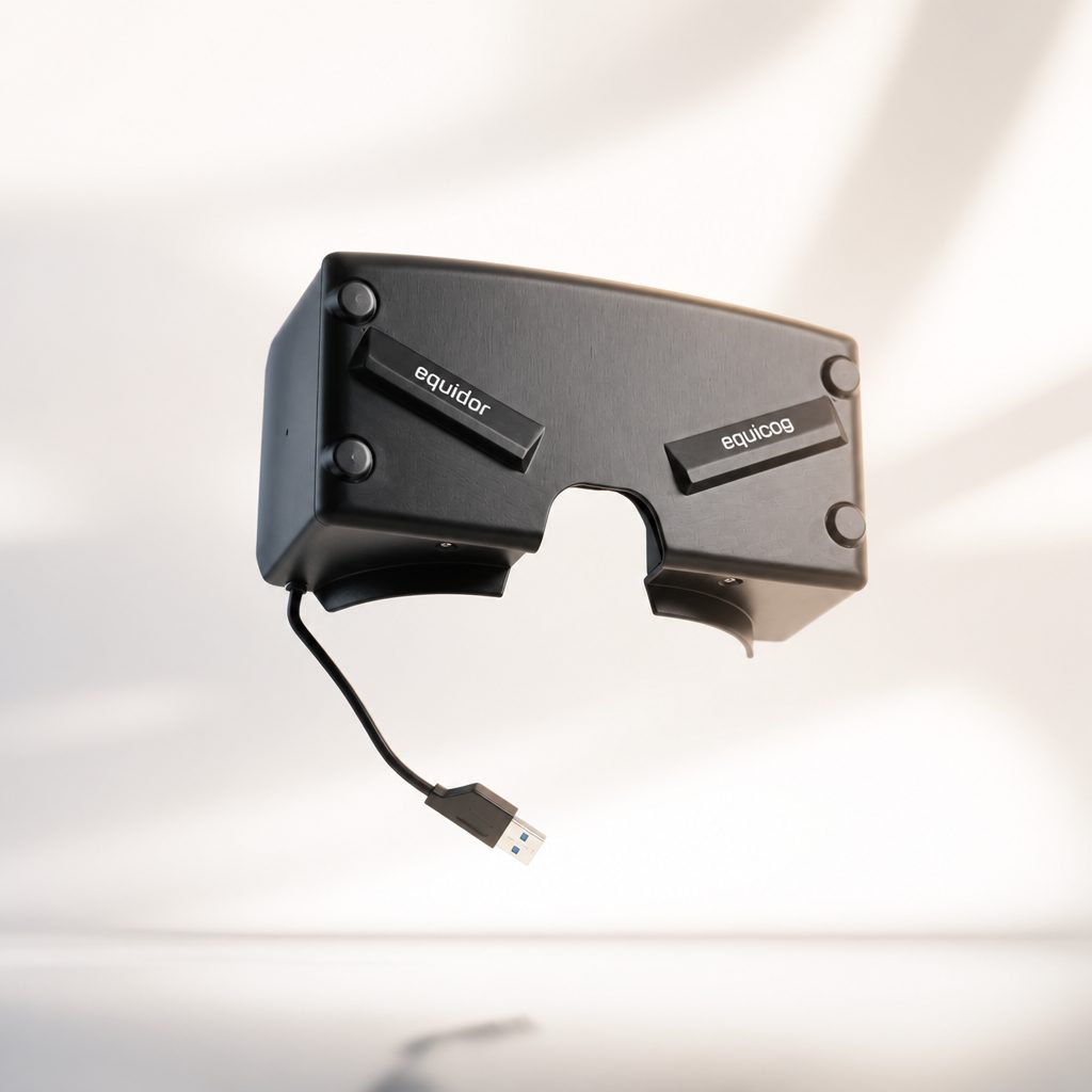
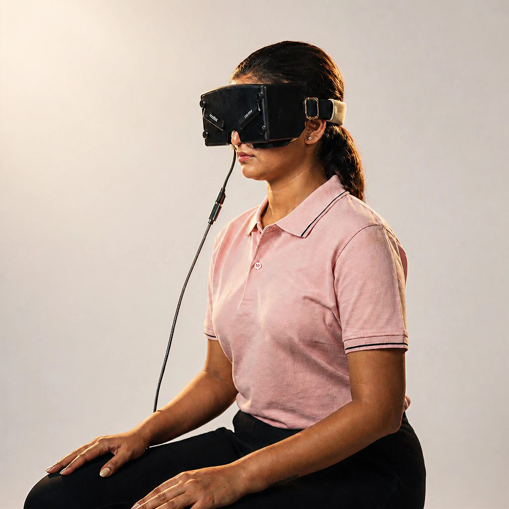
The design choices reinforce that clinical intent. A non-obtrusive in-frame camera layout keeps the patient’s field of view clear — no dichroic IR mirrors, no visor-mounted LEDs with exposed contacts — while each eye is recorded at 100 fps for true high-resolution oculography. Head velocity is captured continuously and replayed as a synchronised head animation alongside the eye video, automatic pupil detection removes the variability of manual threshold setting, and integrated pupillometry adds an autonomic dimension that conventional VNG cannot offer. Fixation and pupillometry lights are delivered through optical principles for safety and clarity, and the entire system runs off a single USB connection — making equiCOG genuinely point-of-care, and giving the patient the assurance that they have been through a more complete and contextually accurate evaluation.
Key Capabilities
- Binocular direct eye-video capture — no obtrusive IR-reflective glass required
- Lightweight goggle design that allows tests to be performed with ease
- Electronics-free fixation visor for enhanced patient safety
- State-of-the-art eye tracking with auto-detection of blinks
- Automatic measurement of pupil size (in mm)
- Real-time head-position rendering and recording for all protocols
- Automatic marking of head positions in positional tests
- Playback of recorded video in slow motion
Hardware Specifications
Test Protocols
Diagnostic / Therapeutic Protocols
- SaccadesFixed and random frequencies / amplitudes, hemifield and full-field testing.
- Smooth PursuitMultiple frequencies, horizontal and vertical directions.
- OptokineticFour velocities, four directions.
- Gaze (with and without fixation)4 s pre, 10 s eccentric, 4 s post for rebound nystagmus; skew deviation assessment.
- PupillometryDiameter and pupil area vs time.
- CaloricsStandard and ice-cold protocols with automatic correction for initial spontaneous nystagmus.
- Nystagmus protocolsSpontaneous in light/dark, high-frequency head shaking, hyperventilation, Valsalva (glottic / nose-and-mouth), Tullio, vibration.
- Advanced SaccadesSelf-paced, anti-saccade, gap and memory-based saccades.
- Advanced PursuitOblique, circular pursuit, neck torsion, head pursuit, variable and fixed-velocity tracking.
- Clinical Chair RotationVisually-enhanced VOR, vision-denied VOR, VOR suppression, cervico-ocular response.
- Positional TestsDiagnostic and therapeutic protocols with real-time head-position rendering; auto marking of head positions with sequence guide for the user.
- Video FrenzelFor any user-defined examination.
Highlights
- Every nystagmus recording is paired with synchronised head-motion data, so the eye finding is read in the same reference frame the patient actually experienced.
- Continuous head tracking turns any manual rotatory chair into a quantitative VOR-gain measurement — no extra hardware required.
- Built-in pupillometry adds an autonomic dimension to standard oculomotor testing, all from the same recording session.
- Captures every trial at 100 fps, so difficult patients can be examined once and reviewed in slow motion as many times as the diagnosis demands.
- Synchronised eye-and-head playback supports both clinical head-impulse review and trainee teaching on a single timeline.